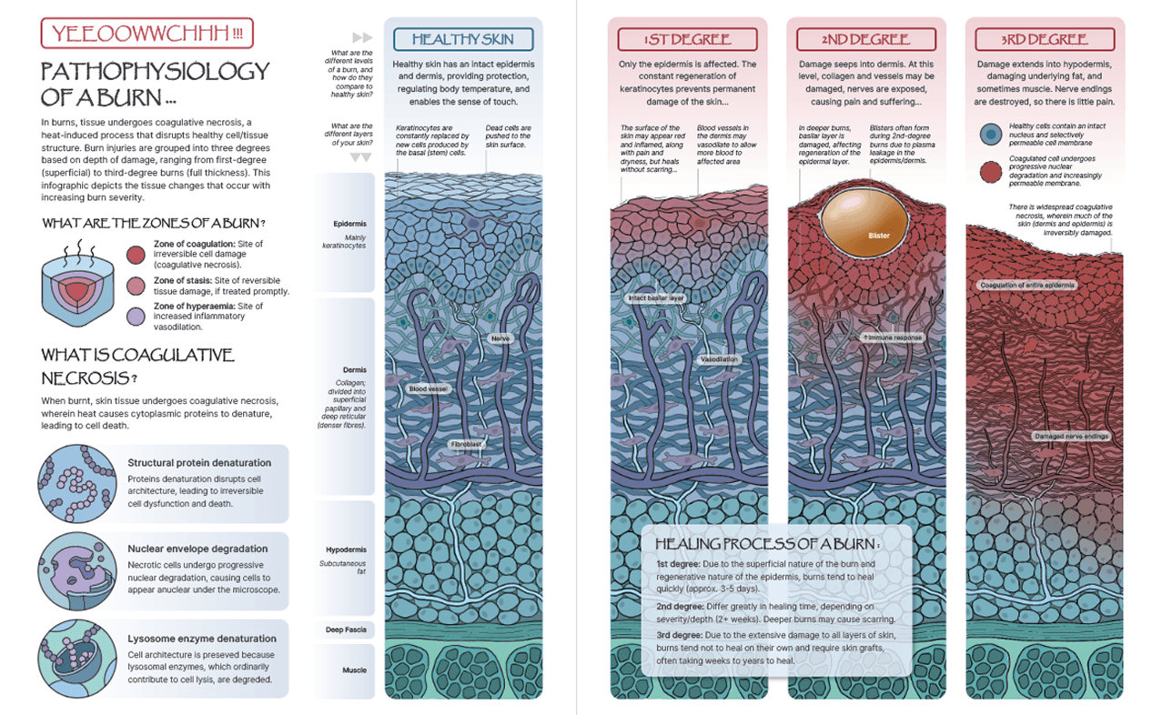
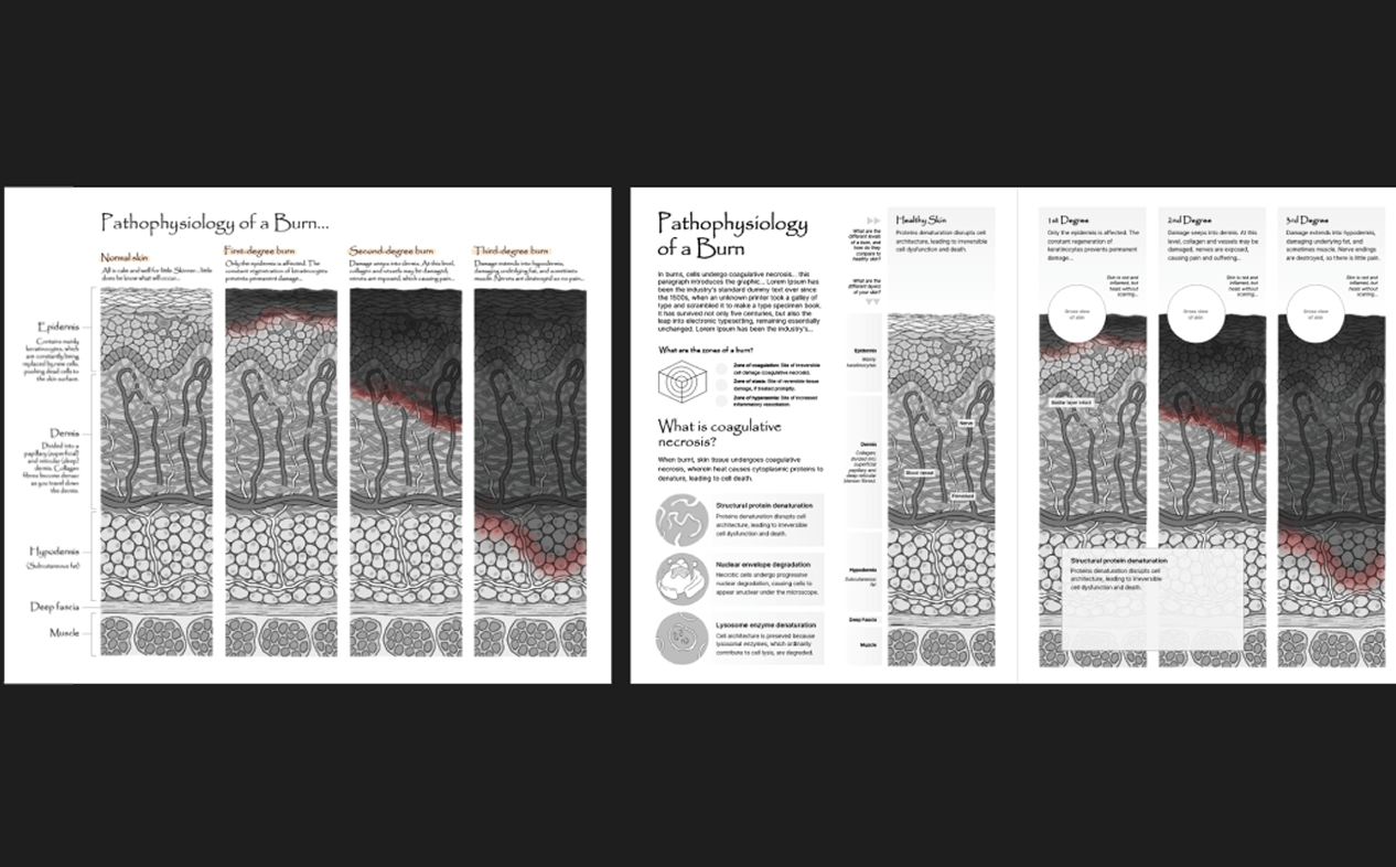
This 2-page conceptual medical illustration spread aims to demonstrate the pathological differences in skin tissue in variably severe burns to an educated lay audience. It uses a flat, contoured style that priortizes clarity over realism.
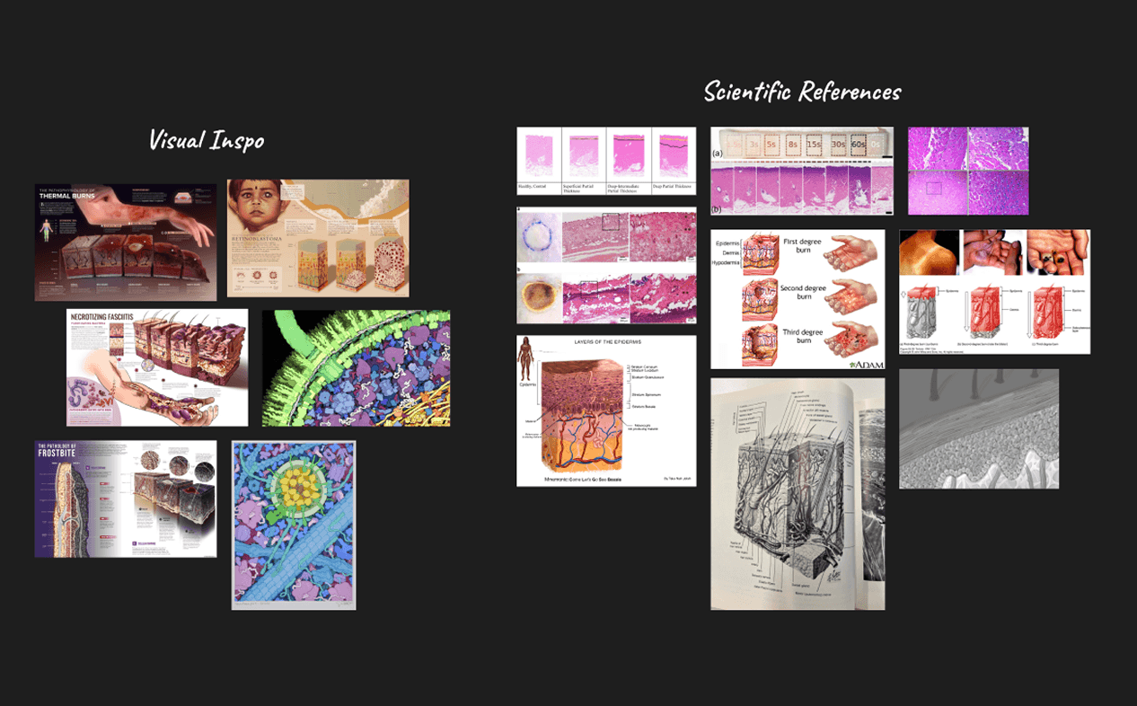
After collecting research on the pathophysiology of thermal burns and consulting existing depictions of burns and coagulative necrosis (e.g. histological slides, illustrations) in books, articles, and atlases, I illustrated a collection of tissue studies of the skin at different levels of burn severity. For these studies, I was inspired greatly by the works of Dr. David Goodsell, aiming for a flat, contoured style that emphasized clarity over realism.
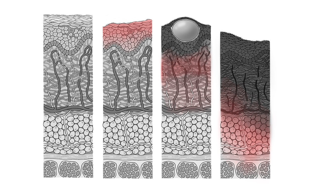
For my comprehensive sketch, I wanted to focus on the tissue studies, making them the largest aspect of my layout so readers could clearly and easily make out the tissue illustrations. I also placed additional sections about the different zones of a burn and the hallmarks of coagulative necrosis to provide more context to readers as they perused the rest of the infographic. For example, the section on lysosome enzyme degradation provides context as to why the cell architecture is still somewhat intact in the right-hand illustrations.
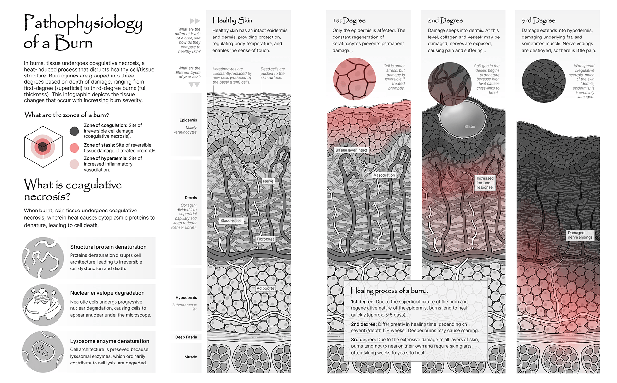
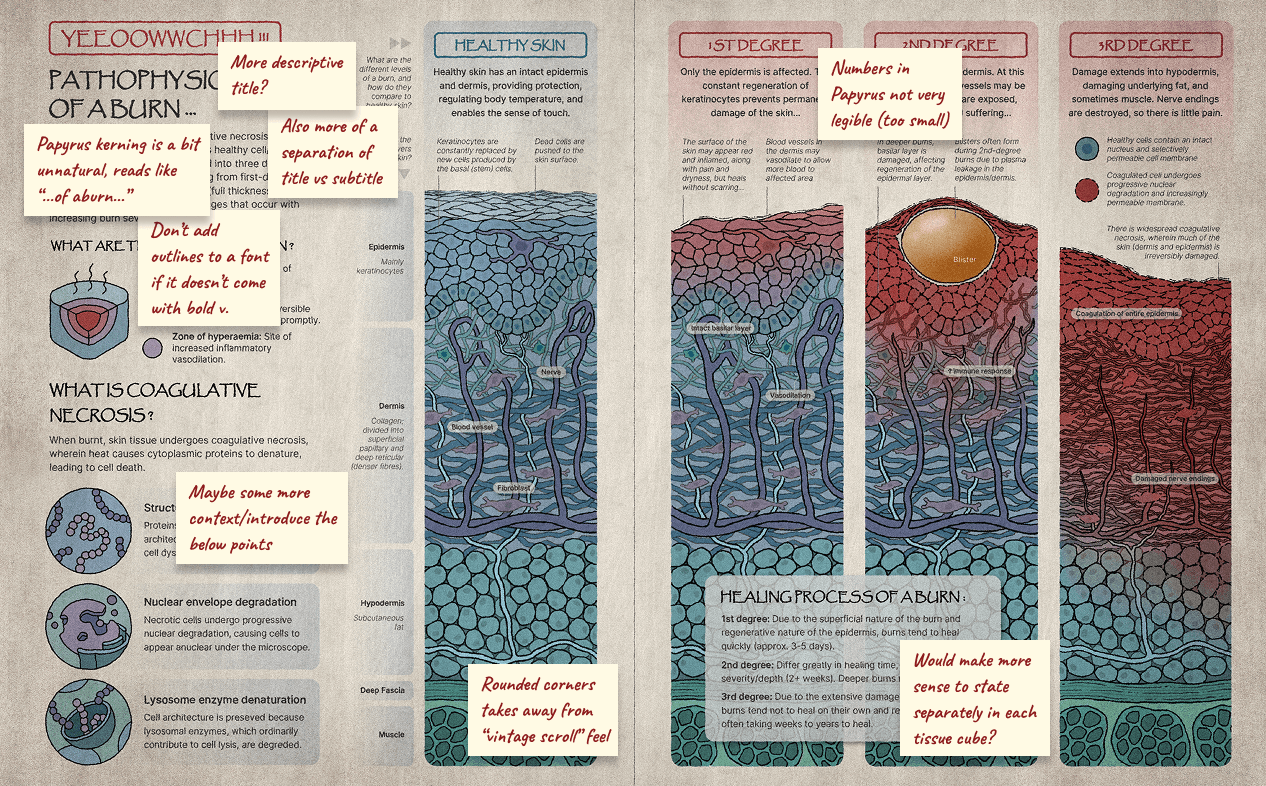
After being reviewed and approved for scientific accuracy by a clinical pathologist, I proceeded to colourize my tissue studies, choosing strong colours to both enhance clarity and give off a slight “watercolour” effect. I also added textures and final touches to the spread in Photoshop for a more vintage aesthetic.