
This didactic illustration was created to visualize the anatomy of the right kidney from an oblique cross-sectional perspective. The goal of this project was to accurately depict the renal structures involved in the production and flow of urine in a lifelike manner, enhancing visual clarity for educational/clinical reference (such as in an atlas).
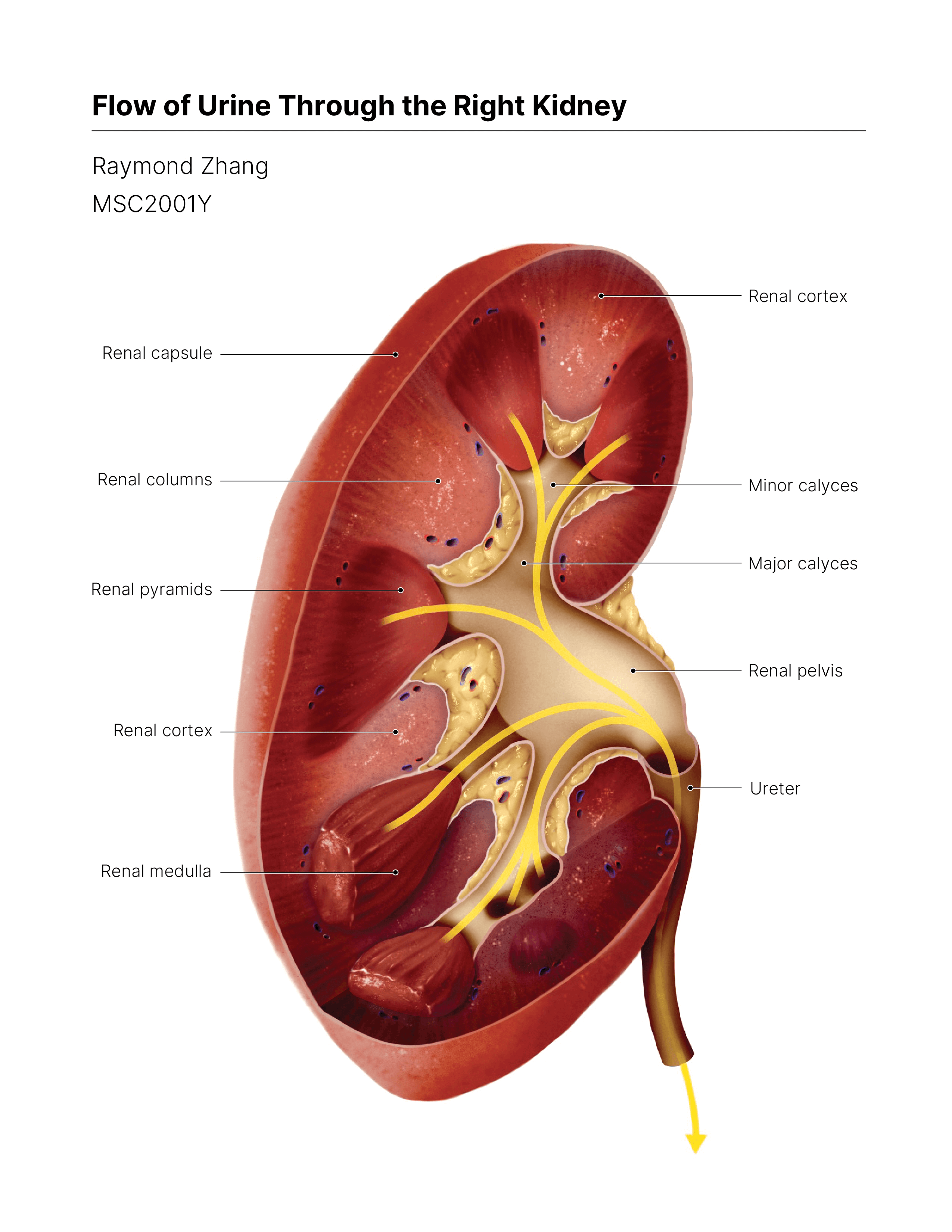
Traditional anatomical illustrations of the kidney often rely on standard sagittal or coronal views, which can limit viewers’ understanding of the spatial relationships between renal structures.
Considerably fewer resources visualize the kidney from an oblique and cross-sectional viewpoint, which can provide a more comprehensive understanding of the overall 3D structure.
To begin, my team and I consulted a range of digital and paper sources that depicted the internal structure of a kidney, including surgical papers, texbooks, and 3D atlases. These materials would provide a base for the maquette and helped inform our design decisions later on, as well as serve as references for texture, form, and colour.
Working off various base models from online 3D databases, we developed a preliminary maquette to serve as a foundation for the final illustration. The models were brought into ZBrush for positioning and texturing, where we added striations, noise, and other organic details to bring the kidney to life. The refined model was then imported into Cinema4D for staging and material application.

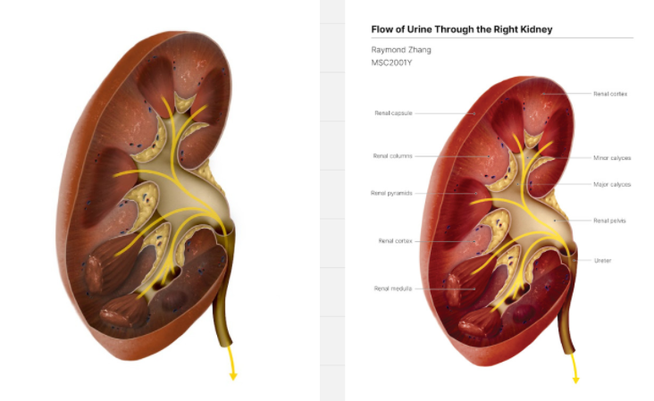
On Procreate, I used the base model to draft a line sketch, adding details such as blood vessels, fat, and arrows. I then blocked out the major shapes of the kidney, which was exported as a .psd file, allowing me to complete the remainder of the illustration in Photoshop. Details were then added using references from the research stage and additional sources. (surgical videos, etc.)
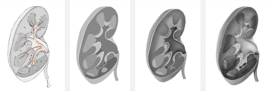
Still working on Photoshop, I used a combination of layer adjustments and blend modes to colourize the grayscale rendering. Labels were added on Illustrator.
- Agur, A. M. R., & Dalley, A. F., II. (2009). Grant's atlas of anatomy. 12th ed. Philadelphia, Wolters Kluwer Health/Lippincott Williams & Wilkins, p. 360.
- Calvo, J. (n.d.). Kidney [Photograph]. Retrieved December 20, 2024 from, https://www.sciencephoto.com/media/1303159/view/kidney
- Calvo, J. (n.d.). Macro section of a normal human kidney [Photograph]. Retrieved December 20, 2024 from, https://www.sciencephoto.com/media/908887/view/macro-section-of-a-normal-human-kidney
- Mahadevan, V. (2019). Anatomy of the kidney and ureter. Surgery (Oxford), 37(7), 359–364. https://doi.org/https://doi.org/10.1016/j.mpsur.2019.04.005
- Moore, K. L., Agur, A. M. R., Dalley, A. F., II., & Moore, K. L. (2015). Essential clinical anatomy. Fifth edition. Philadelphia, Wolters Kluwer Health. p. 314.
- Northwestern Medicine. (2020). Kidney Transplant Surgery | Inside the OR [Video]. YouTube. https://www.youtube.com/watch?v=zPXphpl7gco&ab_channel=NorthwesternMedicine
- Rocco, F., Cozzi, L. A., & Cozzi, G. (2015). Study of the renal segmental arterial anatomy with contrast-enhanced multi-detector computed tomography. Surgical and Radiologic Anatomy, 37(5), 517–526. https://doi.org/10.1007/s00276-014-1382-7
- Rohen, J. & Yokochi, C. (1983). Color Atlas of Anatomy. Igaku-Shoin, p. 306.