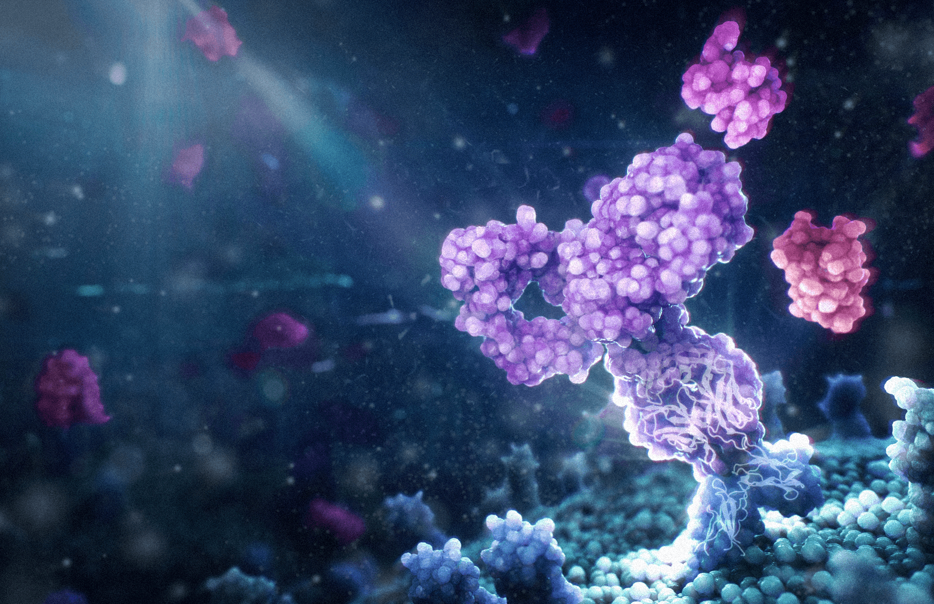
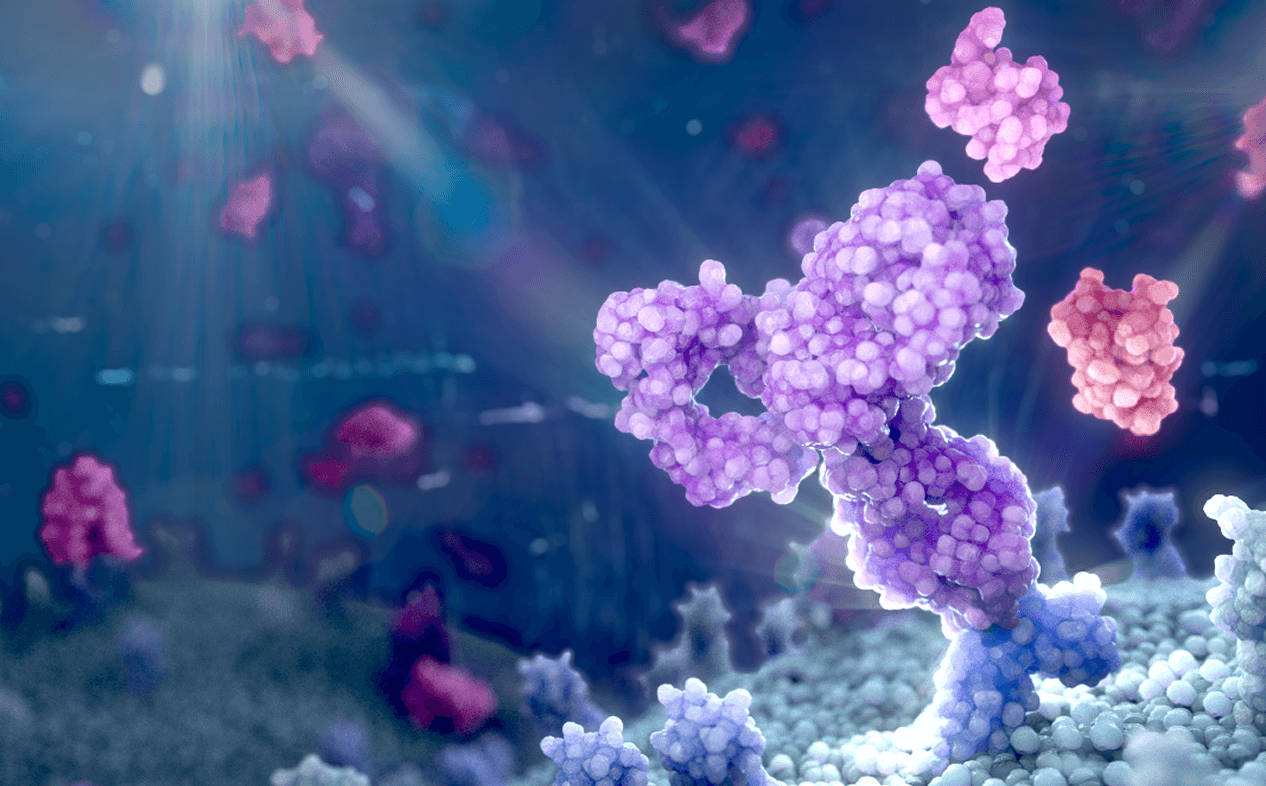
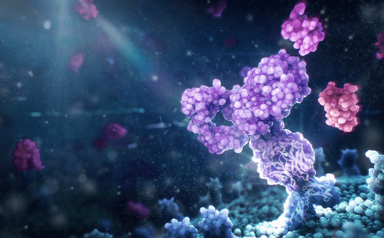
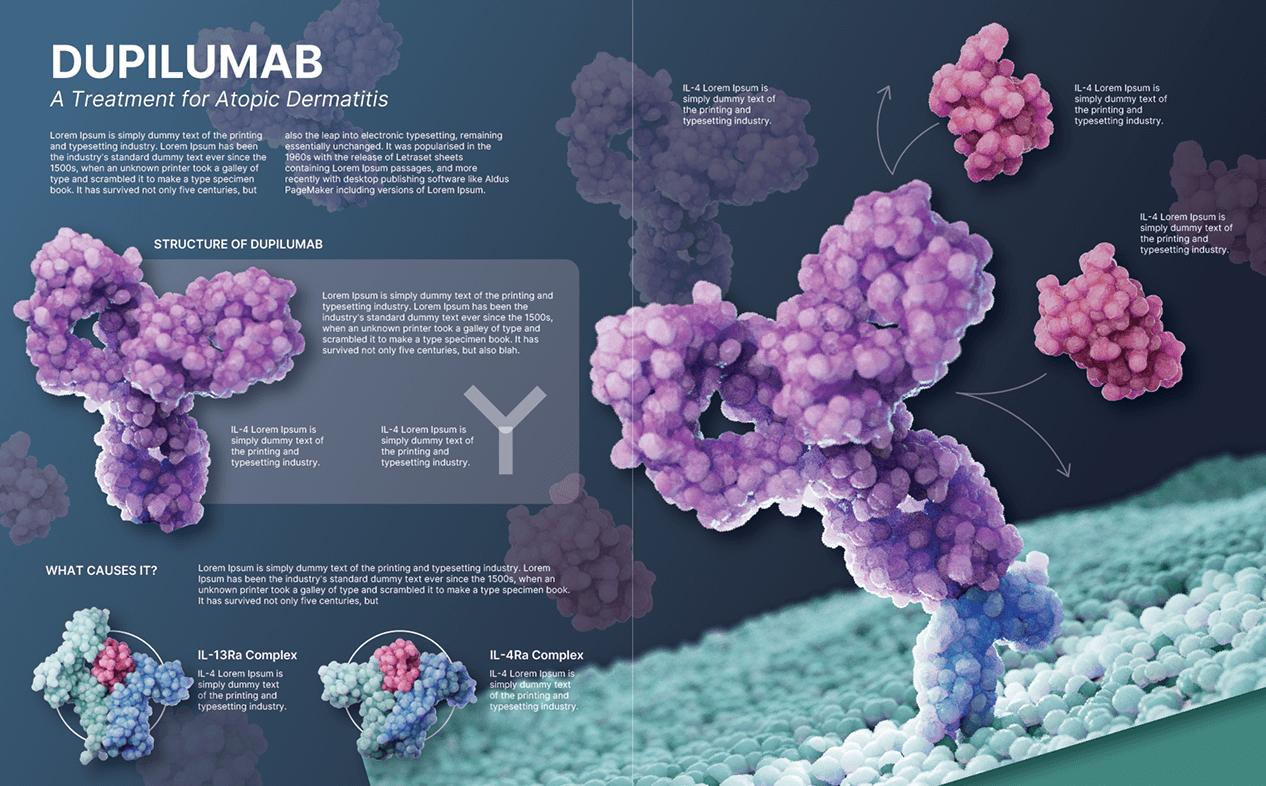
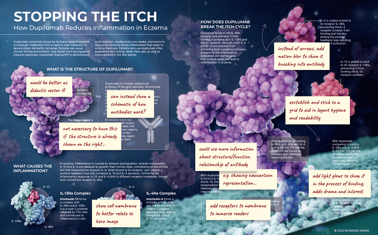
This 2-page magazine spread depicts the structure and mechanism of action of dupilumab, a biologic medication that is used to treat moderate to severe atopic dermatitis (eczema). The focus of the infographic is a molecular visualization of dupilumab binding to its key receptor: IL-4Rα.
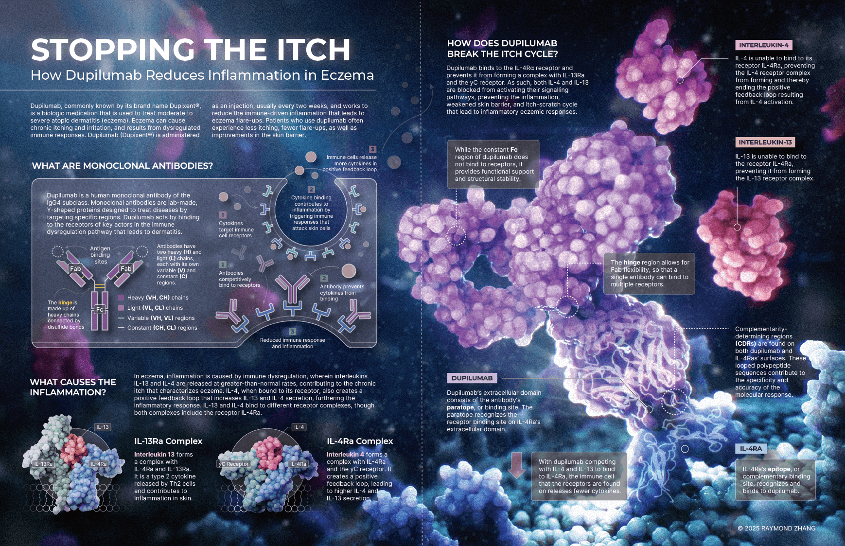
Dupilumab (brand-named Dupixent), is an injectable biologic that reduces inflammation in patients with moderate to severe atopic dermatitis (eczema).
I was curious to understand the cell biology and molecular science behind dupilumab. Unfortunately, there were very few sources online about the structure and function of dupilumab, and even fewer that depicted its mechanism of action in a visual medium.
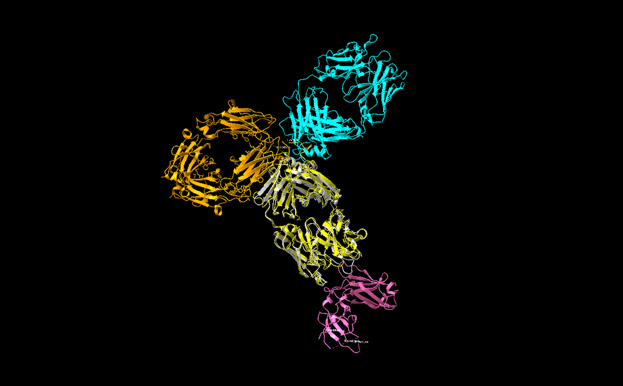
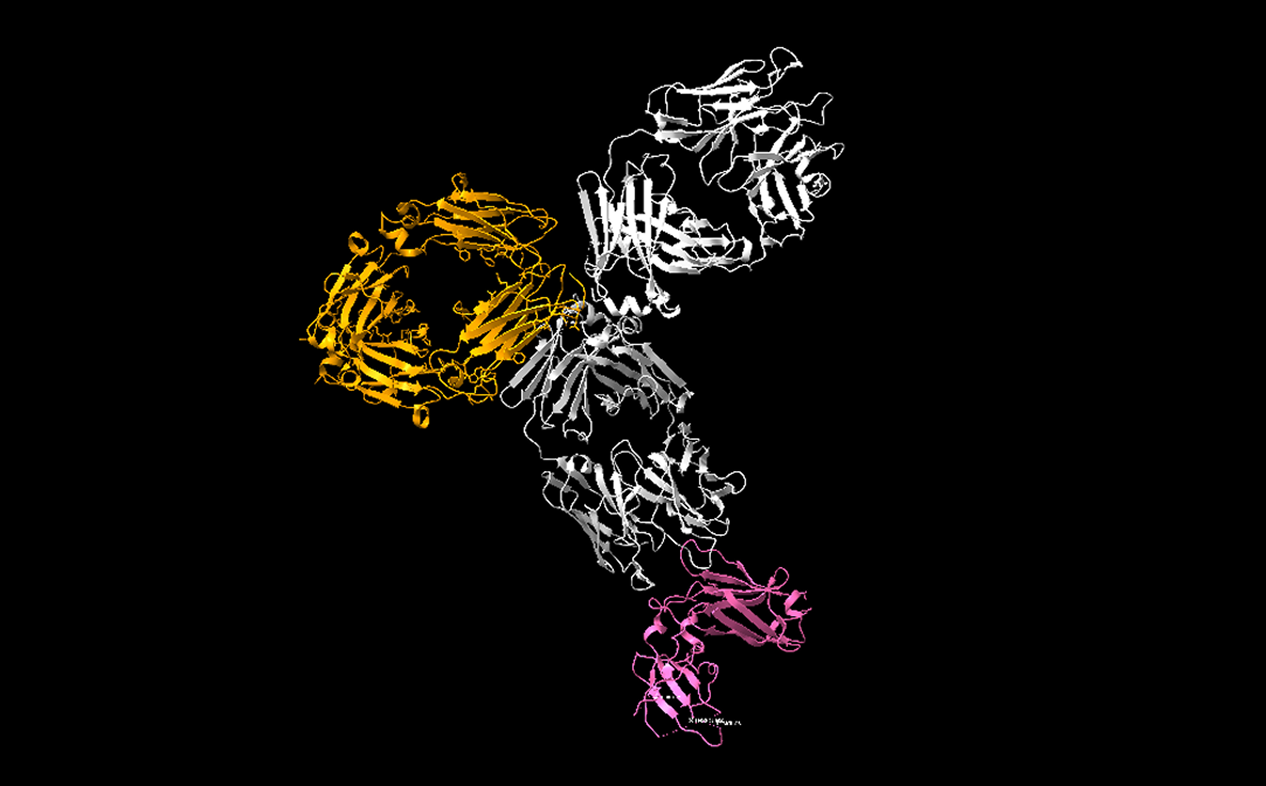
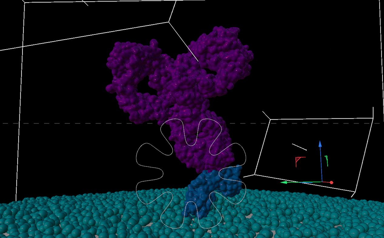
In fact, the full structure of dupilumab is not available publicly, which meant I needed to reference and combine multiple PDB files to create a faithful reconstruction of the molecule. I referred to the RCSB PDB database to gather the molecular structures needed for the project.
Dupilumab is a human monoclonal antibody of the IgG4 subclass. Only the Fab region of dupilumab is available publicly on the RCSB website, so I had to obtain the Fc region from another full-length IgG4 antibody (pembrolizumab). Fortunately, the Fc region is constant within antibody subclasses, so using another IgG4 antibody as the base of the Fc region would still provide me with a faithful representation of dupilumab’s full structure.
After collecting three PDB files to reconstruct the molecule:
- 6WGB: Crystal structure of the Fab portion of dupilumab
- 5DK3: Crystal Structure of Pembrolizumab, a full length IgG4 antibody
- 6WGL: Dupilumab Fab with Crystal Kappa design complexed with human IL-4 receptor
I then imported the files into ChimeraX, where I cleaned up the molecular models and used the Matchmaker tool to splice in the Fab of dupilumab into the pembrolizumab file. In the end, I was left with a reconstruction of dupilumab, which I then exported for modelling/staging in Cinema4D.
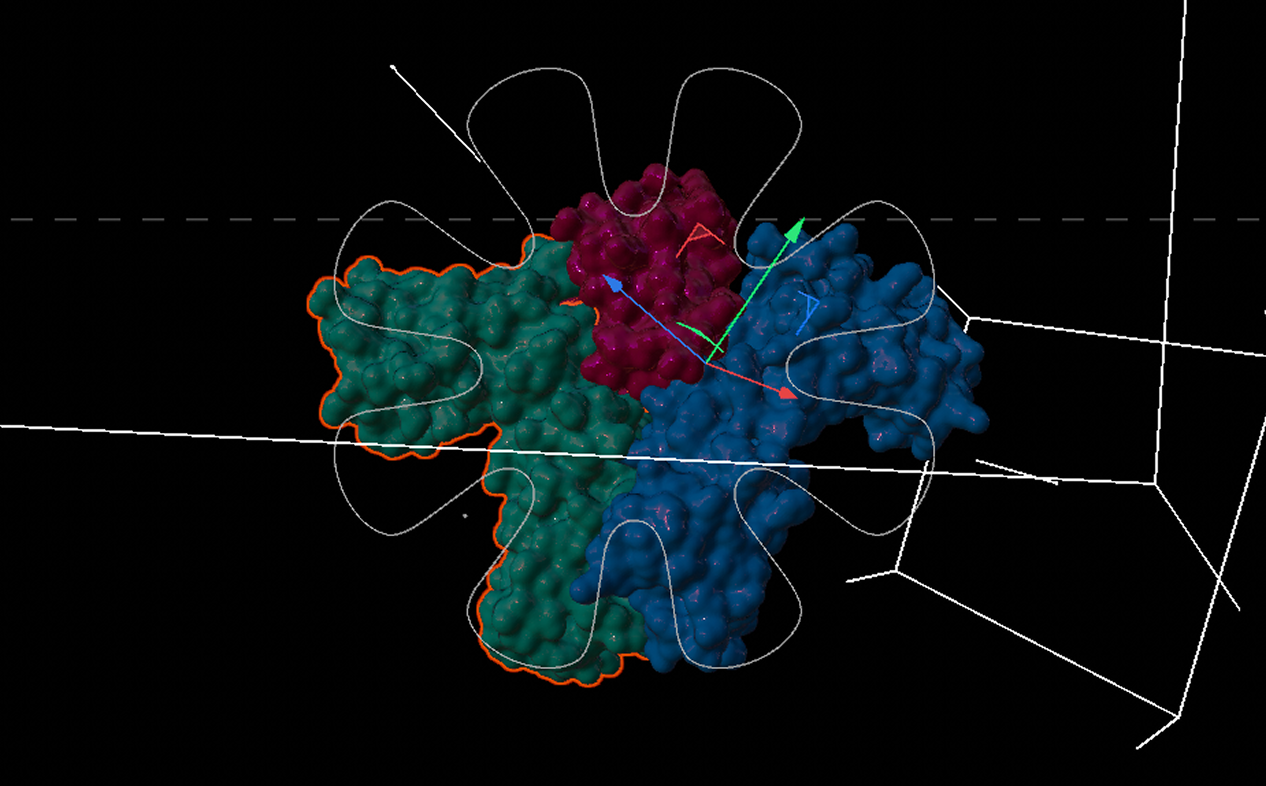
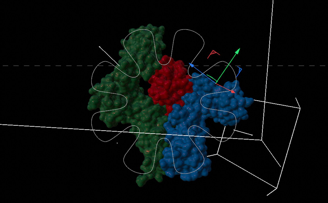
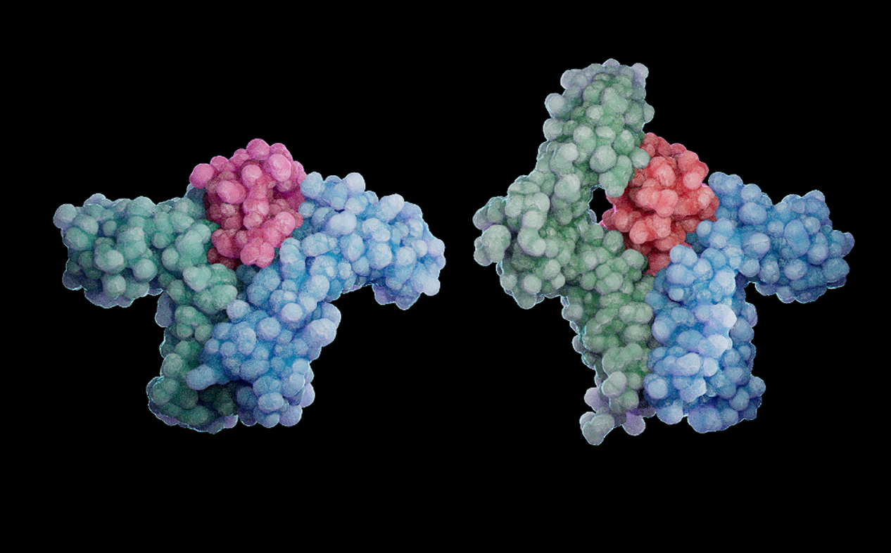
For my infographic, I also needed the structures of interleukin 4 (IL-4) and interleukin 13 (IL-13), as well as their respective receptor complexes. To this end, I collected PDB files of the interleukin-receptor complexes, meaning I only needed two files:
- 3BPL: Crystal structure of the IL4-IL4R-Common Gamma ternary complex
- 3BPO: Crystal structure of the IL13-IL4R-IL13Ra ternary complex
Later, I set up the scene in Cinema4D, using a variety of procedural tools to create an immersive molecular scene that included, dupilumab molecules, cytokines, receptors, and other elements.
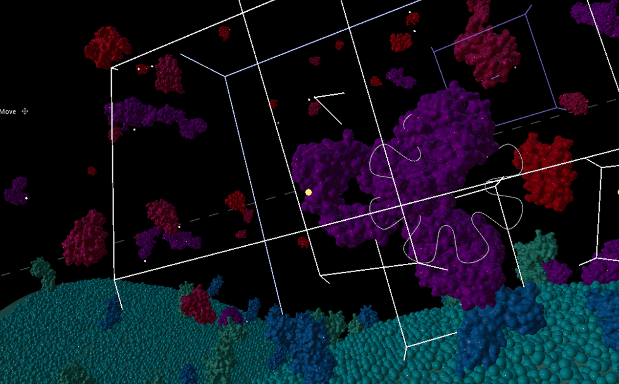
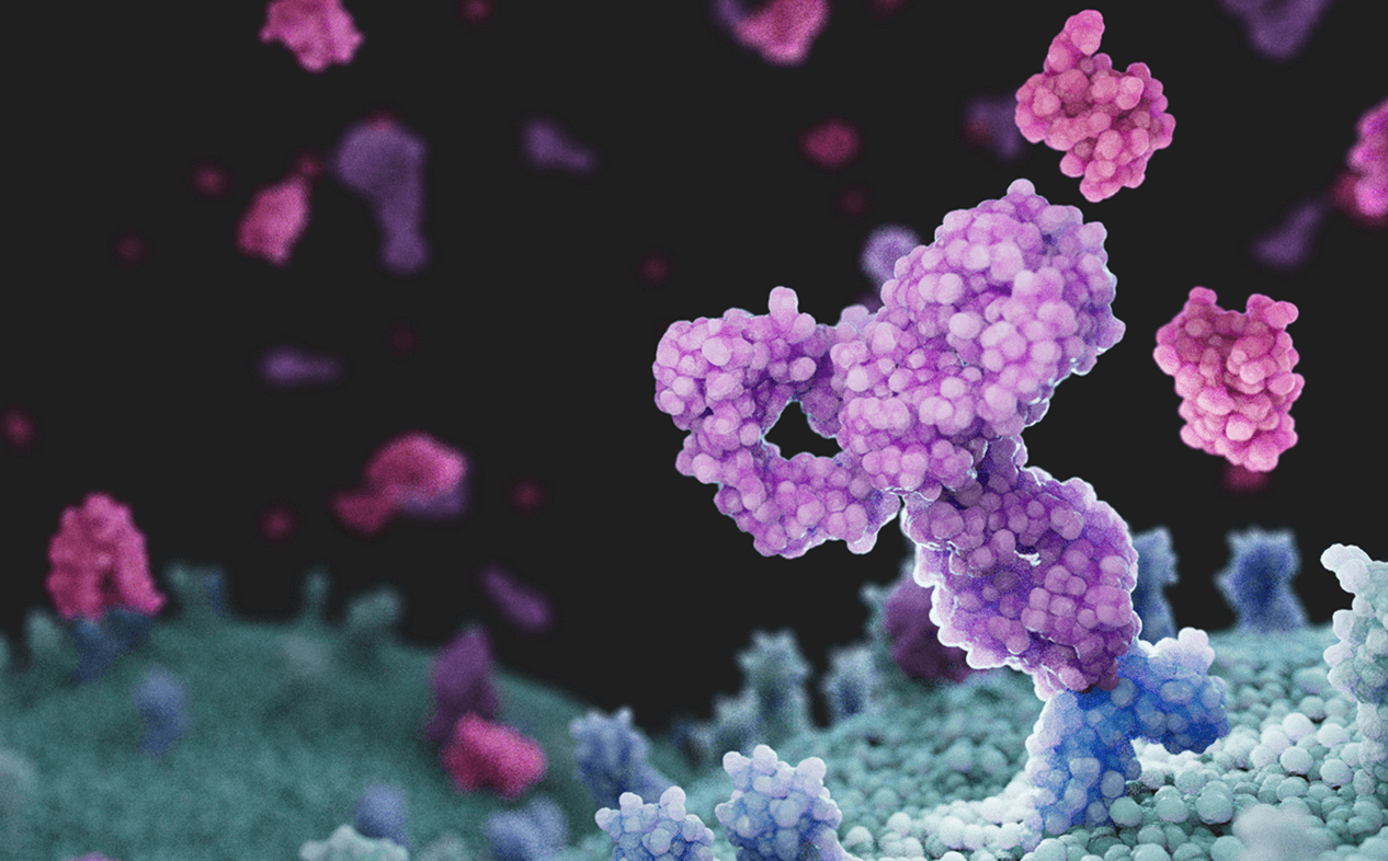
Using the .exr file exported from Cinema4D, I added a background, further lighting, lens flares, and other effects in Adobe After Effects. I chose After Effects due to the many features it offered that could enhance my 3D render, including Maxon’s Red Giant add-on. I experimented with camera effects, chromatic aberration, particle systems, motion blur and other features to create an immersive and realistic molecular environment.
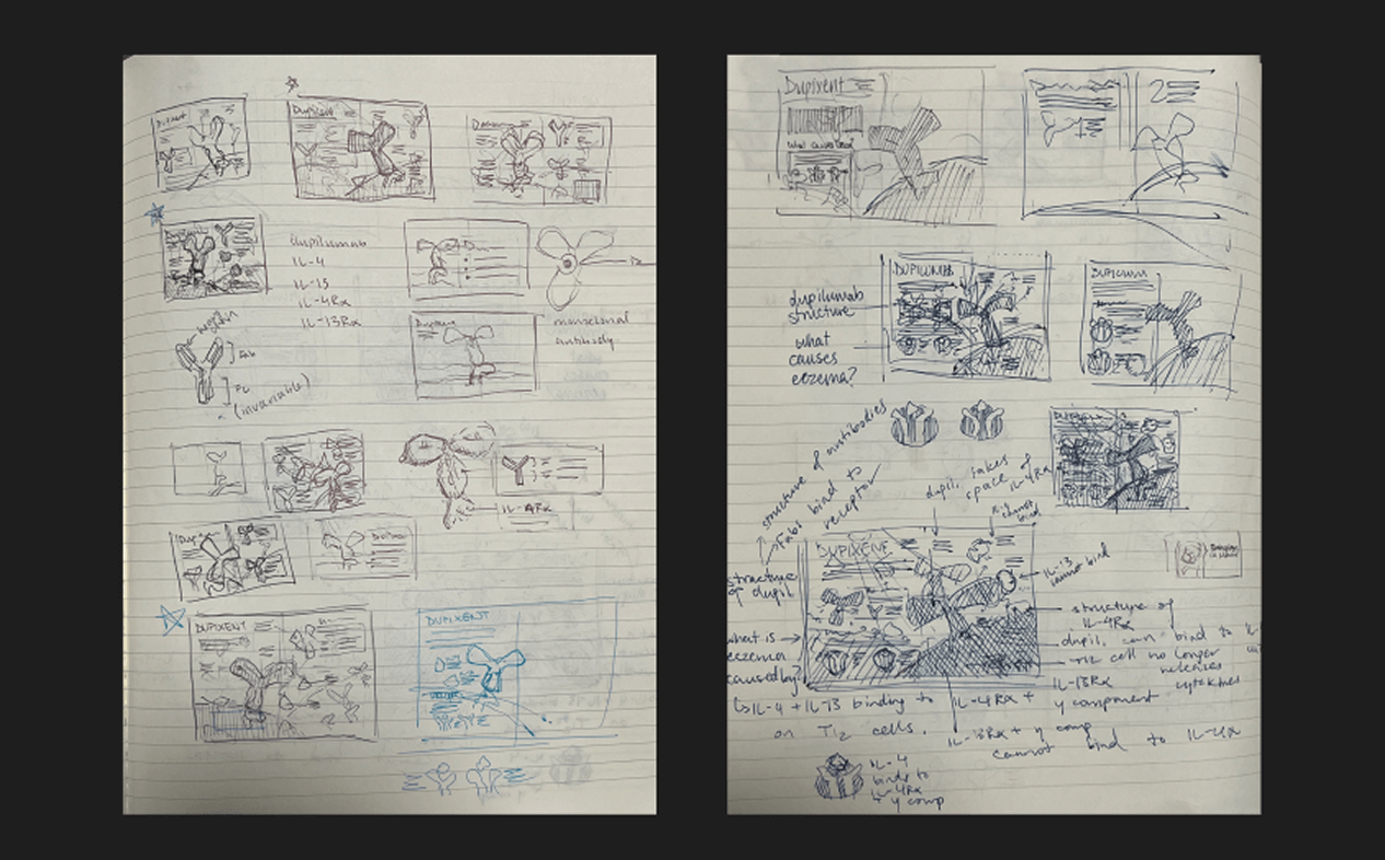
In the very early stages of this project, I created quick thumbnails and layout ideation sketches to inform the placement of elements in my infographic. Later on, I referenced the sketches to create quick concept mock-ups using early renders of my molecular models, which I used to both gather initial feedback from faculty/peers and to provide inspiration for the environment, mood, and overall look and feel of the project.
- Harb H, Chatila TA. Mechanisms of Dupilumab. Clin Exp Allergy. 2020 Jan;50(1):5-14.
- RCSB Protein Data Bank. (2008). 3BPL: Crystal structure of the IL4-IL4R-Common Gamma ternary complex. https://www.rcsb.org/structure/3BPL.
- RCSB Protein Data Bank. (2008). 3BPO: Crystal structure of the IL13-IL4R-IL13Ra ternary complex. https://www.rcsb.org/structure/3BPO.
- RCSB Protein Data Bank. (2015). 5DK3: Crystal Structure of Pembrolizumab, a full length IgG4 antibody. https://www.rcsb.org/structure/5DK3.
- RCSB Protein Data Bank. (2020). 6WGB: Crystal structure of the fab portion of dupilumab. https://www.rcsb.org/structure/6WGB.
- RCSB Protein Data Bank. (2020). 6WGL: Dupilumab Fab with Crystal Kappa design complexed with human IL-4 receptor. https://www.rcsb.org/structure/6WGL.
- Sanofi and Regeneron Pharmaceuticals, Inc. (2025). DUPIXENT® (dupilumab) Mechanism of Action for Uncontrolled Moderate-to-Severe Atopic Dermatitis. https://www.dupixenthcp.com/atopicdermatitis/about/mechanism-of-action.
- Thibodeaux Q. et. al. (2019). A review of dupilumab in the treatment of atopic diseases. Hum Vaccin Immunother, 15(9), 2129–2139.